TORN MENISCUS

ANATOMY AND MECHANISM

Torn Meniscus

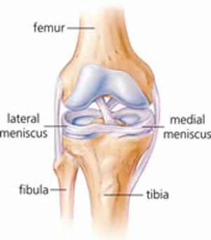
A meniscus is a C-shaped piece of cartilage that acts as a shock absorber of the knee, protecting the bones from wear and tear. There are 2 menisci in each knee joint, 1 one either side. Meniscal tears usually occur as a result of twisting movements, where the foot remains planted on the ground, and are common in sports such as volleyball, netball, basketball, soccer and rugby.

SIGNS AND SYMPTOMS

  • Pain, particularly with activities that involve twisting or bending the knee
  • Clicking and/or locking
  • Swelling – usually 24 hours following injury
  • Difficulty bending and fully straightening knee


The extent of the signs and symptoms will depend on the severity of the tear. With larger tears generally more pain is felt. Also, with certain types of tears, flaps can lift up and cause the knee to lock which is usually very painful.

TREATMENT

Immediate treatment, and within the first 3 days, consists of the RICE principles:

Rest – Rest your knee, avoid too much walking and refrain from playing sport.

Ice  – Every 2 hours apply ice, this will help with the pain and swelling.

If using an ice pack (or frozen peas), leave it on for 10-20 minutes, using a tea towel or similar between the pack and skin to prevent ice burn.

If using an ice cube, “massage” your knee with the ice, gently rubbing it over the skin. Keep the ice cube moving to prevent ice burn and do this for no longer than 5 minutes.

Compression – Use Tubigrip (or a stretchy bandage) on the knee to apply some compression to the joint. This will help to alleviate swelling.

Elevation – Prop your leg up so that your knee is elevated above the level of your heart. This will help to minimise the swelling.

Initial physiotherapy treatment will include gentle joint range of motion and strength exercises aimed at preserving movement and muscle strength. It will also focus on reducing swelling, managing pain and restoring a normal walking pattern. You should do light pedalling on an exercycle with little resistance as this will keep your knee moving with minimal to no pain as it is not having to support your bodyweight.

As pain and swelling decreases your physio will progress your exercise programme, eventually introducing agility and impact exercises. The last phase of rehab will be focusing on sports-specific exercises (if the aim is return to sport).

May 5, 2014 / by /
PLANTAR FASCIOPATHY / ARCH PAIN

ANATOMY, MECHANISM AND SIGNS & SYMPTOMS

Plantar Fasciopathy

The plantar fascia is a thick sheet of connective tissue (ligament) that supports the arch of the foot. It attaches from the heel bone to the base of the toes. There is a fat pad under the heel where the plantar fascia attaches, and this helps to dampen the load through that insertion point.

Plantar fasciopathy is a general term that describes heel or arch pain as a result of an overuse problem of the plantar fascia.

Initially, in the few days following the original injury, this is inflammatory in nature (plantar fasciitis) and symptoms include intense pain (particularly at the end of the day), heat and swelling. The inflammatory phase usually lasts a few days, unless repetitive loading and overuse of the plantar fascia continue, in which case it may last longer.

If repetitive stress of the plantar fascia continues degeneration usually occurs, including calcification of the plantar fascia near the insertion at the heel. In this case the condition is no longer inflammatory. Pain is likely to be worse when putting weight on your foot after a period of rest (e.g. in the morning) and usually eases after the first few steps.

TREATMENT

Treatment will likely include:

  • RICE after injury (Rest, Ice, Compression, Elevation)
  • Strapping
  • Soft orthotics
  • Use of night splints
  • Stretches
  • Exercises
  • Ice
  • Massage
  • Replacing footwear if necessary
  • Acupuncture
  • Activity modification
May 5, 2014 / by /
LATERAL ANKLE SPRAIN

ANATOMY & MECHANISM

The most common type of sprain to the ankle occurs when the foot rolls inwards. When this happens, one or more of the ligaments on the outside of the ankle get stretched or, if the force is great enough, tear.

There are 3 ligaments on the outside of your ankle, called the anterior talofibular ligament, posterior talofibular ligament and the calcaneofibular ligament:

Lateral Ankle Sprain

This type of injury, called an inversion ankle sprain, is common  in sports such as netball, basketball, soccer and running. Simply stepping off a curb awkwardly or twisting your foot on uneven ground can also result in an inversion ankle sprain.

SIGNS AND SYMPTOMS

  • Pain and swelling around the outside of the foot
  • Difficulty and pain with walking
  • Pain particularly when turning the foot in

The extent of the signs and symptoms will depend on the severity of the sprain and the number of ligaments involved.

Grade 1 – Only a few of the ligament fibers are torn

Grade 2 – Roughly 50% of the ligament fibers are torn

Grade 3 – The ligament has ruptured (torn through completely)

TREATMENT

Immediate treatment, and within the first 3 days, consists of the RICE principles:

Rest – rest the ankle, avoid too much walking and refrain from playing sport.

Ice  – Every 2 hours apply ice, this will help with the pain and swelling.

If using an ice pack (or frozen peas), leave it on for 10-20 minutes, using a tea towel or similar between the pack and skin to prevent ice burn.

If using an ice cube, “massage” the outside of your ankle with the ice, gently rubbing it over the skin. Keep the ice cube moving to prevent ice burn and do this for no longer than 5 minutes.

Compression – Use an ankle brace or slightly tight sock, or even better Tubigrip, on the ankle to apply some compression, this will help to alleviate the swelling.

Elevation – Prop your leg up so that your ankle is elevated above the level of your heart. This will help to minimise the swelling.

From there, physiotherapy treatment will start with gentle movement, strengthening and balance exercises. Once the ligaments have healed sufficiently more vigorous exercises, such as agility and sport specific training. Your physiotherapist may recommend using an ankle brace when returning to sport, either initially or for ongoing use. This will depend on the individual and the nature of the injury.

May 5, 2014 / by /
ACL RUPTURE

ANATOMY AND MECHANISM

ACL Rupture

The anterior cruciate ligament (ACL) of the knee attaches to the back of the femur (thigh bone) and front of the tibia (shin bone) and is one of the main stabilisers of the knee. It prevents excessive movement of the tibia forward in relation to the femur, and also provides some rotational stability.

It is commonly injured in sports such as netball and rugby, when stopping suddenly. For example being tackled in rugby, when the lower leg is suddenly immobilised and the knee hyper-extends.

The ACL may be strained (only some of the fibers torn) or ruptured (all of the fibers torn).

SIGNS AND SYMPTOMS

  • Pain
  • Instability (feeling that the knee may “give way”)
  • Immediate severe swelling
  • Audible “pop” sound at time of injury (with a ruptured ACL)
  • Difficulty walking
  • Limited range of movement

TREATMENT

Immediate treatment, and within the first 3 days, consists of the RICE principles:

Rest – rest the knee, avoid too much walking and refrain from playing sport.

Ice  – Every 2 hours apply ice, this will help with the pain and swelling.

If using an ice pack (or frozen peas), leave it on for 10-20 minutes, using a tea towel or similar between the pack and skin to prevent ice burn.

If using an ice cube, “massage” your knee with the ice, gently rubbing it over the skin. Keep the ice cube moving to prevent ice burn and do this for no longer than 5 minutes.

Compression – Use Tubigrip if you have some on the knee to apply some compression, this will help to alleviate the swelling.

Elevation – Prop your leg up so that your knee is elevated above the level of your heart. This will help to minimise the swelling.

If the ACL is strained treatment will likely be conservative (non-surgical). If the ACL is ruptured treatment may include surgical repair of the ligament or conservative treatment.

Surgery is often recommended to athletes who intend to continue competing in demanding sport. This involves a reconstruction of the ligament rather than a repair, where a graft is taken from a hamstrings tendon or the patellar tendon and replaces the ruptured tendon. This is done arthroscopically (key-hole surgery).

Following surgery it will be at least 6-9 months before you can return to sport and you will require physiotherapy rehabilitation. This will initially include exercises to return the joint’s normal range of motion, followed by a strengthening programme which is gradually progressed to introduce more stressful/demanding activities and, finally, a functional programme aimed at sport-specific activities to aim for returning to playing sport.

If you do not have surgery, physiotherapy rehabilitation will still be important. This will consist of exercises to retain the joint’s range of motion, reduce swelling and increase strength of the muscles around the knee in order to increase stability. You may still be able to play some sport, but this will need to be discussed with your physiotherapist and/or specialist.

May 5, 2014 / by /
Carpal Tunnel Syndrome

Carpal tunnel syndrome develops when a large nerve becomes compressed in the wrist. This is the nerve that controls the feeling to thumb, index finger and thumb side of the ring finger. This nerve also controls the muscles at the base of the thumb.The Carpal Tunnel is a small tunnel that allows this nerve and the tendons to the fingers to pass through. Anything making this tunnel smaller (such as inflammation of the tendons) results in compression on the nerve diminishing circulation and causing pain, numbness, and tingling in fingers.

It can be caused by:

  • Wrist injury or fracture to the wrist/forearm.
  • Frequent use of vibrating tools.
  • Repetitive motion with a bent wrist (typing).


Symptoms include:

  • Numbness, burning, tingling or pain in hand and fingers.
  • Increased discomfort at night and in the morning
  • Discomfort with prolonged wrist flexion.
  • Weakness in the hand.


Treatment from the Physiotherapist will be centred on attempting to decrease inflammation in the wrist and minimise aggravating activities.

Splints and tape can be used to correct movements and aid in the resting period of the injury.

An exercise programme can aid in improving the symptoms, combined with nerve glides has shown excellent results for treating carpal tunnel syndrome.

April 17, 2014 / by /
Lateral Epicondylalgia (Tennis Elbow)

Tennis elbow is the common name for pain radiating from the outside of the elbow. It is most often caused by overuse of the forearm muscles causing inflammation of the tendons (such as in tennis).It is suspected that small tears in the tendon cause tennis elbow from overuse – these begin to heal but are re-injured by continued use. This injured tendon becomes fixed in a state of degeneration similar to scar tissue.The symptoms of tennis elbow are;

  • Difficulty holing onto, picking up or griping objects.
  • Pain, stiffness and/or decreased elbow and hand movement.
  • Forearm and muscle tightness.
  • Insufficient functional strength
  • Point tenderness around the outside of the elbow.


Treatment involves:

  • An initial focus on the R.I.C.E protocol, resting and icing to help minimise inflammation and pain.
  • If the condition is long standing the Physiotherapist may employ modalities to help increase blood supply and stimulate healing.
  • Exercises focussing on the eccentric action of movement, hoping to both strengthen and correct the alignment of the internal fibres.
  • Stretching is also given as a means to correct the alignment of fibres.
April 17, 2014 / by /
Acromioclavicular (AC) Joint Sprain

The A.C. Joint is a combination of the collarbone and shoulder blade and it forms the top of the shoulder joint. In this joint there are ligaments holding the bones together similar to other joints, and as such can be injured in a similar manner.

The most common cause of injury to this joint is to fall onto the shoulder or with the arm outstretched. There are many levels of severity when it comes to the AC joint, the most common of which is a sprain. An AC joint sprain is characterised by:

  • Tenderness over the top of the shoulder, localised to one spot
  • Pain with movement (especially reaching across the body)


Assessment will involve clearing all other structures that could be involved including the neck, and other areas of the shoulder. Once the AC joint sprain has be identified and diagnosed the following is likely to be included in treatment.

  • Rest, Ice, Compression, Elevation to minimise initial damage
  • Slow and steady strengthening program to build up the surrounding muscle
  • Stretches to ensure the ligaments heal in the correct orientation
  • More functional strengthening to get you back to where you need to be.
April 17, 2014 / by /
Adhesive Capsulitis or ‘Frozen Shoulder’

Although the exact mechanism causing frozen shoulder is unknown it is very common post-trauma, following injury and surgery. It can occur after injury to neural or muscular structures, or in some cases can occur spontaneously. It is most common between 40-60 year olds and more so in women. Although frozen shoulder often resolves on it’s own, un-aided it can take between 12 – 30months.Frozen shoulder is characterised by a systematic process in which the shoulder becomes less painful albeit more stiff causing trouble with undressing and reaching behind.A typical Physiotherapy assessment will involve observing movement, testing strength, and range of motion. The Physiotherapist will assess the ability of your nerves to glide to rule out all other shoulder problems.With the assessment combined with the list and timeline of symptoms your physiotherapist will be able to give a well-reasoned diagnosis.


Treatment will consist of some of the following:

  • Stretches of various muscle groups to loosen the surrounding muscles
  • Massage in order to release surrounding tissue
  • Strengthening exercises to decrease the load on the shoulder joint
  • Hands on modalities to stretch the joint capsule and help restore movement.

Research shows that a physiotherapy programme can help in shortening the period of frozen shoulder and also aid in a more complete recovery.

April 17, 2014 / by /
Rotator Cuff Problems

The rotator cuff is made up of four muscles of which encompass the shoulder. They act to stabilise and provide rotational movement of the arm. When these muscles become inflamed they can become injured and painful.The shoulder joint is a ball in socket type joint made up of the arm, and shoulder blade. It is the rotator cuff that holds these two together.  Inflammation often occurs in sports requiring the arm to be moved over the head repetitively, for example; tennis, swimming and weight lifting.This group of muscles can further be injured through trauma in which the shoulder is forced into an awkward position or from falling on to an outstretched hand.Symptoms of a rotator cuff injury include:

  • Pain with arm movement; in particularly with overhead activities.
  • Pain at night, especially lying on the injured side.
  • Weakness with raising arm above the head and difficulty with activities such as brushing hair, reaching for objects etc.
  • A loss of range of motion of shoulder.


The Physiotherapist will most likely assess your strength, range of motion and palpate around the shoulder region. This will include trying to provoke the symptoms that you are having at home. The Physio will look and check for pain with resistance and for the signs there could be narrowing of the shoulder space causing chronic inflammation.

Treatment involves addressing both the current symptoms but also the underlying biomechanical deficiency that lead to the injury.  The initial treatment is to rest from aggravating activities and icing the region to reduce and control the inflammation and pain.

Our Physiotherapist will prescribe a strengthening programme aimed at rehabilitating the injured muscle and attending to the causative factors such as poor posture.

April 17, 2014 / by /